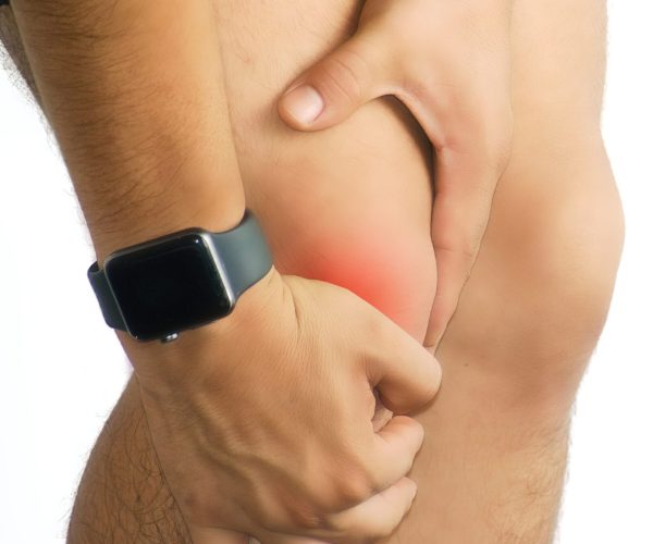
Knee Dislocation: Diagnosis, Treatment & Recovery

What is a Knee Dislocation?
A knee dislocation is a serious injury where the tibia (shinbone) and femur (thighbone) lose their normal alignment—often called a tibiofemoral dislocation.
Unlike kneecap subluxation, this involves displacement of the main joint and is a medical emergency due to potential vascular and nerve damage.
Although rare—affecting about 1 in 100,000 people annually—many cases go unreported because 50% self-reduce before patients reach the hospital. Knee dislocation accounts for only 0.001–0.013% of all orthopaedic injuries, with a male-to-female ratio of 4:1.
Causes & Risk Factors
These injuries most often arise from high-energy trauma, including:
- Vehicle collisions (“dashboard injuries”)
- Falls from heights
- Crush or athletic trauma
However, low-energy dislocations can occur in severely obese individuals or during routine activities.
Knee dislocations are categorised by the direction of tibial displacement relative to the femur:
- Anterior (~30–50%): from hyperextension
- Posterior (~30–40%): from axial loading to a bent knee
- Less common: medial, lateral, and rotatory


Signs, Symptoms & Urgency
Common Signs:
- Intense pain and obvious deformity; knee may appear dislocated or misshapen
- Inability to bear weight or move the leg
Critical Warning Signs:
- Compromised leg blood flow, risking ischemia and amputation if untreated within 8 hours
- Injured popliteal artery in up to 40% of high-energy cases
- Nerve damage, especially to the common peroneal nerve (≈20% incidence), leading to foot drop between 15%–35%
- Associated injuries: fractures (~60%), ligament tears (ACL, PCL, MCL, LCL), meniscal or tendon damage
Diagnosis & Assessment
1.Clinical Evaluation
- Detailed injury history, mechanism, initial deformity, and post-injury changes.
- Physical exam includes inspection, palpation, range of motion, neurovascular assessment (distal pulses, capillary refill, sensation, motor), and ligament integrity tests.
2.Imaging
- X-rays before and after reduction confirm alignment and detect fractures.
- CT angiography if the ankle-brachial index is <0.9 or pulses are abnormal to assess blood vessels .
- MRI to evaluate ligamentous, cartilage, and soft tissue injury after stabilization

Emergency Management & Initial Care
- Immediate reduction is typically performed in a controlled setting with sedation to restore joint alignment
- Once aligned, a rigid splint or external fixator may be used to stabilise the knee if it is unstable
- Urgent assessment for vascular compromise is critical—absent pulses or ischemic signs require emergency vascular surgery.
- Evaluate for nerve, ligament, tendon, meniscus, and compartment syndrome involvement.

Definitive Treatment: Surgery & Rehabilitation
Surgical Timing:
- Early multiligament reconstruction (ideally within 3–4 weeks) is preferred for optimal function and stability
- One-stage reconstruction of injured ligaments (ACL, PCL, collateral, posterolateral corner) yields better outcomes than staged procedures
Repair Techniques:
- Cruciate ligaments repaired or reconstructed using grafts (autografts/allografts).
- Collateral ligaments repaired or augmented as needed.
- ACL/PCL/PLC often require combined grafting procedures for strong stability.
Postoperative Care:
- Begin physiotherapy immediately to regain mobility, aiming for 0–90° range by day one.
- Rehabilitation protocols often last 9–15 months and focus on gradual strengthening and functional recovery.
- Reassessment for residual nerve or artery damage is essential.
Long-Term Outlook
- Recovery varies: low-velocity injuries generally have better outcomes; high-velocity trauma may lead to lingering stiffness, nerve impairment, arthritis, or vascular issues .
- 10% risk of amputation, associated with delayed vascular reconstruction beyond 8 hours.
- Close postoperative care and structured rehabilitation are crucial for restoring joint strength and function.
Preventive Measures for Knee Dislocation
While not all knee dislocations can be prevented—especially those caused by high-impact trauma—there are several proactive steps individuals can take to reduce the risk of injury or recurrence.
Strengthening the muscles that stabilise the knee, particularly the quadriceps, hamstrings, and hip abductors, can significantly improve joint stability. Engaging in regular balance and proprioception training helps the body respond more effectively to sudden movements, decreasing the chance of awkward landings or twisting injuries.
Proper warm-up routines and flexibility exercises before sports or physical activity enhance joint readiness and reduce strain. For those with a history of instability or anatomical risk factors, using supportive braces or taping techniques during high-risk activities may offer additional protection.
Finally, maintaining a healthy body weight lowers stress on the knee joint, further minimizing injury risk.

Why Choose Clifford Orthopaedics?
At The Clifford Clinic, we prioritise safety and outcomes through:
- 24/7 orthopaedic trauma access and expedited care pathways
- Vascular and nerve evaluation, including swift CT angiography
- Expertise in multiligament reconstruction, including advanced PCL and PLC repairs
- Custom rehabilitation programs led by skilled physiotherapists
- Long-term support for monitoring recovery, adjusting rehab, and minimising long-term complications
Frequently Asked Questions
What is the difference between a knee dislocation and a kneecap (patella) dislocation?
A knee dislocation (tibiofemoral dislocation) involves the displacement of the entire joint between the femur (thigh bone) and tibia (shin bone). This is a rare but severe injury that often damages multiple ligaments and can disrupt blood vessels or nerves.
In contrast, a patellar dislocation only affects the kneecap, which slips out of its groove on the femur (usually to the side) but leaves the main knee joint intact. While both can be painful and limit mobility, tibiofemoral dislocations are more serious and require urgent medical evaluation due to the risk of vascular or neurological damage.
How do I know if a knee dislocation has affected blood vessels or nerves?
Vascular and nerve injuries are among the most dangerous complications of knee dislocations. Signs of vascular injury may include:
A cold or pale foot
Absent or weak pulse in the foot (especially the dorsalis pedis or posterior tibial pulse)
Numbness or tingling
Severe swelling or bruising
Nerve damage, particularly to the common peroneal nerve, may present as:
Numbness or burning sensation on the outer shin or top of the foot
Inability to lift the foot (foot drop)
Weakness in ankle or toe movements
These signs warrant immediate emergency care. A delay in restoring blood flow can lead to permanent damage or even amputation.
Is surgery always required for a knee dislocation?
Not always—but in most cases, especially with multiligament injuries (involving ACL, PCL, MCL, LCL, or posterolateral corner), surgery is typically recommended. Minor dislocations without significant ligament or vascular damage may be managed conservatively with reduction, bracing, and physical therapy. However, if the joint remains unstable, or if MRI shows major soft tissue damage, surgical reconstruction is often needed to restore long-term joint function and prevent early arthritis or recurrent dislocations.
What does recovery and rehabilitation after knee dislocation surgery involve?
Rehabilitation is a long-term process and often spans 9 to 15 months. It begins with early range-of-motion exercises (typically starting within a few days post-op), progressing gradually to strengthening and proprioceptive training.
Milestones include:
- Regaining 0–90° knee motion within the first few weeks
- Partial weight-bearing with support (brace/crutches) in early stages
- Full weight-bearing and functional retraining over several months
A structured rehab program is essential to restore stability, flexibility, and strength. Long-term outcomes are generally better when physiotherapy is closely guided by your orthopedic team.
Can a knee dislocation cause permanent disability or complications?
Yes, especially if not treated promptly. Complications include:
- Chronic instability or stiffness from ligament damage
- Early onset post-traumatic arthritis
- Permanent nerve damage, such as foot drop
- Compartment syndrome, a rare but limb-threatening condition
- Amputation, in extreme cases where vascular supply is compromised and not restored in time
That’s why timely diagnosis, vascular assessment, and orthopedic intervention are critical. With proper treatment, many patients can regain good function, but ongoing monitoring and rehabilitation are essential.
Our Services
EFFECTIVE SOLUTIONS
Don’t let knee osteoarthritis hold you back.
Start with a clinical assessment today—and get a tailored plan to manage pain, improve function, and preserve quality of life for years to come.


