Patellar Tendinopathy (Jumper’s Knee):
A Comprehensive Guide
What Is Patellar Tendinopathy?
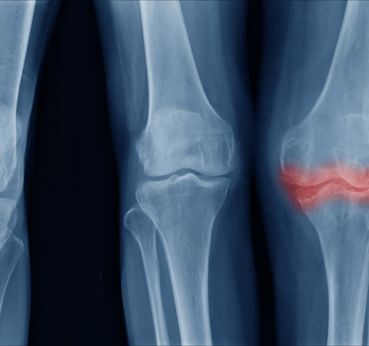
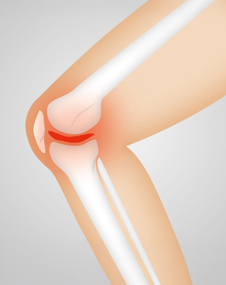
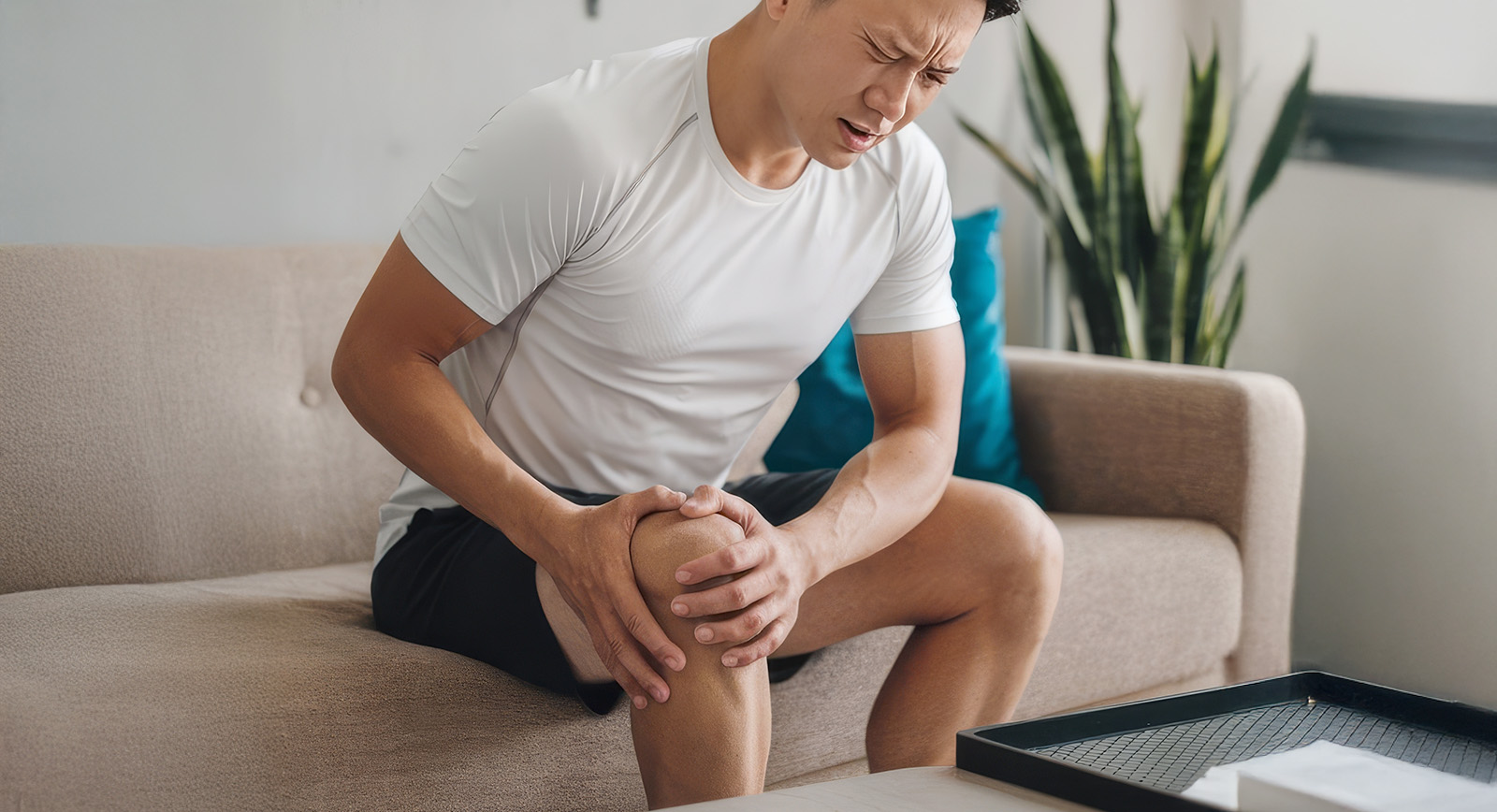
Patellar tendinopathy, often called jumper’s knee, is a condition affecting the tendon that connects your kneecap (patella) to your shin bone (tibia). This tendon acts as a critical link in your knee extensor mechanism—transferring the force generated by your quadriceps muscle to straighten your knee .
Patellar tendinopathy can manifest in two primary forms:
Tendonitis: inflammation of the tendon tissue
Tendinosis: microscopic degeneration and microtears in the tendon, without significant inflammation
It typically occurs at the tendon’s lower patellar attachment (inferior pole) and is particularly common among athletes involved in jumping and running sports
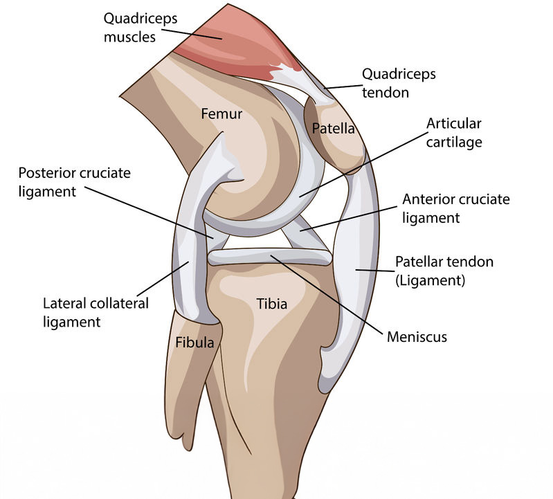
Anatomy of the Patellar Tendon
The patellar tendon emerges as a continuation of the quadriceps tendon, passing beneath the kneecap before attaching to the tibial tuberosity. This arrangement acts like a spring, storing and releasing energy during powerful movements such as jumping and landing.
Key structures involved:
Quadriceps muscle group: generates force to extend the knee
Patella (kneecap): improves leverage and force transmission
Patellar tendon: transfers force to the shin
Infrapatellar bursae and Hoffa’s fat pad: help reduce friction and cushion underlying bone structures
Regain joint comfort today—book your consultation with Clifford Orthopaedics.
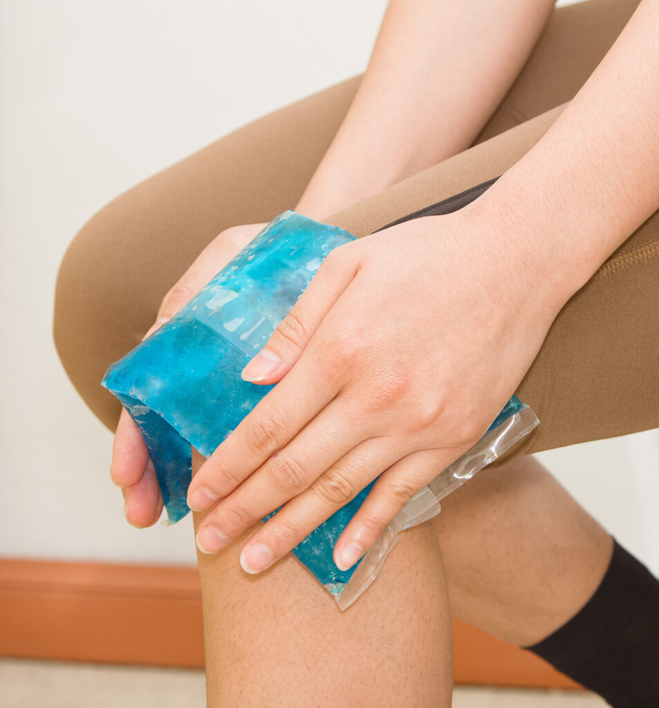
RICE stands for Rest, Ice, Compression, and Elevation—a simple and effective way to manage pain and swelling in knee conditions like patellar tendinopathy.
Rest
Avoid activities that strain the knee, such as jumping or squatting. Use relative rest—gentle movement is okay, but avoid anything painful.
Ice
Apply an ice pack to the front of the knee for 15–20 minutes every 2–3 hours, especially during flare-ups. This helps reduce swelling and numbs pain. Always wrap the ice—don’t apply directly to skin.
Compression
Use a knee sleeve or elastic bandage to reduce swelling and support the joint. Make sure it’s snug but not too tight.
Elevation
Prop your leg up on pillows so your knee is above heart level. This helps drain fluid and reduce pressure in the joint.
RegenPRP® Therapy for Patellar Tendinopathy
When conservative treatments like RICE and physiotherapy don’t provide lasting relief, RegenPRP® (Platelet-Rich Plasma) offers a powerful regenerative solution.
RegenPRP® is an advanced therapy that uses your own blood to promote natural tendon healing. A small blood sample is drawn and processed using a Swiss-engineered RegenKit® system to extract platelet-rich plasma, which is rich in growth factors.
These growth factors:
Stimulate tissue repair
Encourage new collagen production
Reduce inflammation at the tendon site
💉 How Is It Done?
Step 1: Your blood is drawn and spun in a special centrifuge.
Step 2: The concentrated PRP is injected precisely into the affected tendon under ultrasound guidance.
Step 3: You follow a guided rehab plan to support tendon regeneration.
✅ Benefits of RegenPRP®
Addresses the root cause (tendon degeneration), not just symptoms
Minimally invasive and done in-clinic
Uses your own cells—no synthetic drugs or risk of allergy
Proven improvements in pain, stiffness, and function over weeks to months
Suitable for chronic or stubborn tendinopathy unresponsive to rest and physiotherapy.
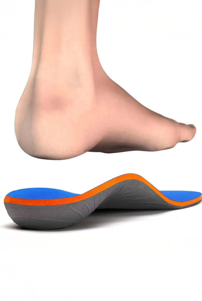
Foot Orthotics: A Customised Solution
Your feet form the foundation of movement, and poor foot alignment can create stress that travels upward—placing extra strain on your knees, hips, and tendons. In patellar tendinopathy, even slight biomechanical imbalances in the feet can worsen tendon loading during walking, running, or jumping.
For example:
Flat feet (overpronation) cause inward knee collapse, increasing tension on the patellar tendon
High arches (supination) reduce shock absorption, concentrating impact on the knees
Uneven leg lengths or abnormal foot angles can shift forces unevenly, overloading one knee more than the other
Reduces tendon overload by improving lower limb biomechanics
May be custom-made or off-the-shelf, based on your needs
👟 Choosing Proper Footwear
Look for shoes that offer:
Good arch support
Shock-absorbing soles
Heel and ankle stability
When Is Surgery Considered?
Surgery becomes an option if symptoms persist despite optimized conservative care for 6–12 months:
Tendon debridement to remove degenerated tissue
Tear repair for partial or complete tendon ruptures Recovery includes immobilization, gradual physiotherapy, and functional rehab.
Why Choose Clifford Orthopaedics?
We deliver comprehensive, evidence-based care tailored to your condition and lifestyle:
Multidisciplinary assessment: orthopaedic evaluation, physiotherapy, imaging support
Individualised rehab planning: carefully staged loading supported by skilled physiotherapists
Access to advanced treatments: extracorporeal shockwave therapy, guided injections when required
Focused on long-term joint health: prevention, education, and movement re-education
Our Services
EFFECTIVE SOLUTIONS
Facing persistent knee pain? Take control of your mobility and comfort—schedule your consultation today and begin your journey towards relief with personalized, expert care.